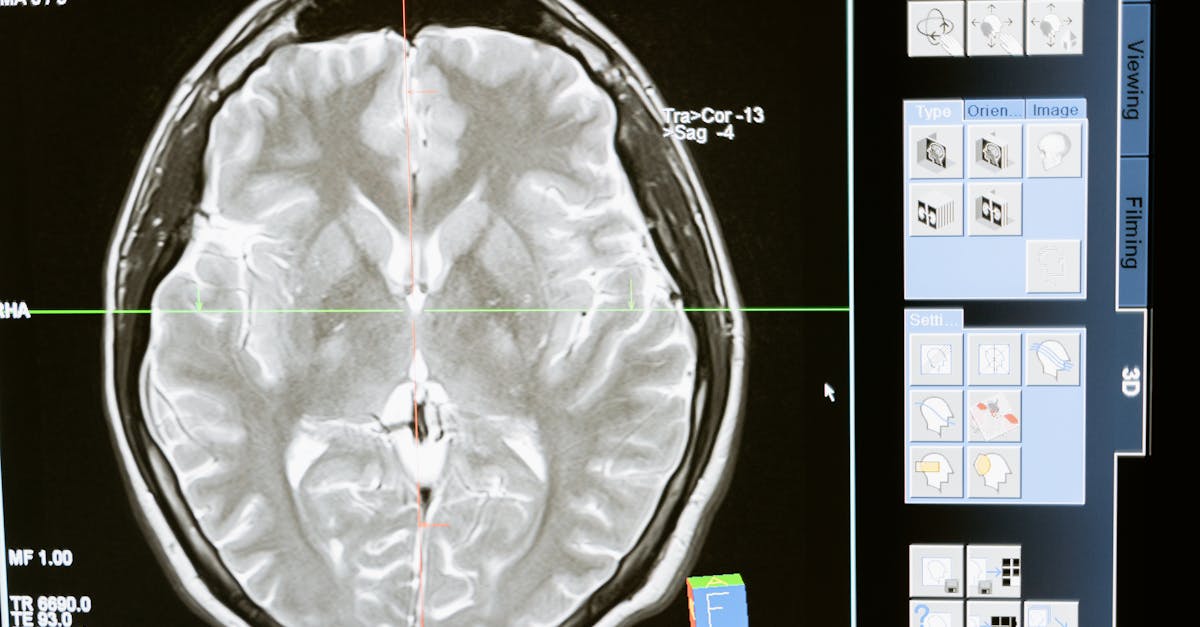
MRCP PACES: Mastering Cranial Nerve Examination Essentials
Why the Cranial Nerve Examination Terrifies PACES Candidates
Let's be honest — the neurology station in MRCP PACES is where many otherwise excellent candidates stumble. And within that station, nothing causes more anxiety than a cranial nerve examination. It demands precision, fluency, a systematic approach, and the ability to interpret subtle findings under the watchful eyes of two examiners who have seen it all.
But here's the truth: the cranial nerve examination is actually a golden opportunity to demonstrate clinical competence. If you can perform it smoothly, identify the key signs, and — critically — localise the lesion accurately, you will score well not just on physical examination but on the discussion that follows, which often carries the most weight.
In this guide, I will walk you through a high-yield, exam-tested cranial nerve examination framework that I have refined through years of teaching PACES candidates, along with the clinical pearls that examiners love to probe.
Understanding What Examiners Are Looking For
Before diving into technique, you need to understand what the examiners are actually assessing. The MRCP PACES mark scheme evaluates four domains across all stations:
Physical Examination — Is your technique correct, systematic, and fluent?
Identifying Physical Signs — Can you accurately elicit and interpret findings?
Clinical Communication — Can you present your findings clearly and logically?
Differential Diagnosis and Clinical Judgement — Can you localise the lesion, formulate a differential, and suggest appropriate investigations?
For the cranial nerve examination, most candidates perform adequately on the technique but fall down on interpretation and localisation. The examiners are not just watching your hands — they are evaluating whether you can think like a physician, not a technician.
The Framework: A Step-by-Step Cranial Nerve Examination
Preparation
Before you even touch the patient, establish your routine:
"I would like to examine this patient's cranial nerves. I have washed my hands, confirmed the patient's identity, obtained consent, and ensured they are comfortable and positioned at 45 degrees."
Begin with a general inspection — take two steps back and observe the patient. Look for:
Facial asymmetry (particularly around the eyes and mouth)
Ptosis (unilateral or bilateral)
Abnormal head posture (head tilt in CN IV palsy, head turn in CN VI palsy)
Wasting of temporalis or masseter muscles (CN V involvement)
Facial scars or previous surgical incisions (e.g., craniotomy, parotidectomy)
Hearing aids or glasses
This 10-second inspection can give you the diagnosis before you lay a hand on the patient.
CN I: Olfactory Nerve
Clinical Pearl: CN I is rarely tested in PACES but may be relevant if the patient has a frontal lobe tumour or post-traumatic presentation. Only test if instructed or if the history suggests olfactory involvement.
Ask the patient: "Have you noticed any change in your sense of smell or taste?"
If clinically indicated, test each nostril separately using non-irritant substances (coffee, vanilla).
CN II: Optic Nerve
This is where many candidates lose marks. The optic nerve examination must include four key components:
1. Visual Acuity
Use a Snellen chart at 6 metres (or a near-vision chart if the patient is bed-bound). Correct for refractive error using a pinhole.
2. Visual Fields
Use confrontation testing with a red pin (5mm target is ideal):
Sit opposite the patient at arm's length.
Test each eye separately (cover the contralateral eye).
Move the target slowly from the periphery to the centre in all four quadrants.
Also test for central scotoma by comparing your vision with the patient's — move the red pin across the midline.
High-Yield Finding: A bitemporal hemianopia suggests a pituitary lesion (chiasmal compression). A homonymous hemianopia suggests a posterior cerebral artery stroke affecting the optic radiation or occipital cortex.
3. Pupillary Reflexes
Test in a dimly lit room (ask the examiners to dim the lights if possible):
Direct and consensual light reflex: Shine a bright light into each eye and observe both pupils.
Swinging light test (for RAPD): Swing the light between the two eyes every 2–3 seconds. Look for paradoxical dilation of the affected pupil when light is swung to it — this indicates a relative afferent pupillary defect (RAPD), pathognomonic of optic nerve disease (e.g., optic neuritis in multiple sclerosis).
Accommodation reflex: Ask the patient to look at a distant target, then at your finger held 30cm away. The pupils should constrict.
Classic PACES Trap: An Argyll Robertson pupil (small, irregular pupils that accommodate but do not react to light) suggests neurosyphilis. A Holmes-Adie pupil (unilateral dilated pupil with slow constriction to accommodation) is benign.
4. Fundoscopy
Perform direct ophthalmoscopy. Key findings to identify:
| Finding | Clinical Significance |
|---|---|
| Swollen optic disc (papilloedema) | Raised intracranial pressure, malignant hypertension |
| Optic atrophy (pale disc) | Previous optic neuritis, compressive lesion, hereditary (e.g., Leber's) |
| Cup-to-disc ratio > 0.5 | Glaucomatous cupping |
CN III, IV, and VI: Oculomotor, Trochlear, and Abducens Nerves
These nerves are tested together because they collectively control eye movements.
Inspection
Look for:
Ptosis (CN III — levator palpebrae superioris)
Squint (strabismus) — note if it is convergent or divergent
Eye position in primary gaze
Pupil Examination
A CN III palsy can be either:
Medical (pupil-sparing): Typically microvascular (diabetes, hypertension). The parasympathetic fibres run peripherally and are spared.
Surgical (pupil-involving): Typically compressive (posterior communicating artery aneurysm, uncal herniation). The peripheral parasympathetic fibres are affected first, causing pupillary dilation.
Examiner's Favourite Question: "Why does a posterior communicating artery aneurysm cause a pupil-involving CN III palsy, but diabetes causes a pupil-sparing palsy?"
Answer: The parasympathetic (pupilloconstrictor) fibres run in the superomedial periphery of the nerve. Compressive lesions (aneurysm) affect these peripheral fibres first, causing early pupillary dilation. Microvascular disease (diabetes) affects the central core of the nerve, sparing the peripheral parasympathetic fibres.
Eye Movements
Ask the patient to follow your finger (or the traditional H-pattern) with their head still:
Horizontal movements: Look right, look left (tests CN VI for abduction, CN III for adduction)
Vertical movements: Look up, look down (tests CN III and IV)
Diagonal movements: Up-right, down-right, up-left, down-left
Ask specifically: "Do you see double at any point?" and note whether the diplopia is horizontal, vertical, or torsional.
Interpreting eye movement findings:
| Nerve | Muscle(s) Affected | Key Findings |
|---|---|---|
| CN III palsy | Medial rectus, superior rectus, inferior rectus, inferior oblique, levator palpebrae | Down and out eye, ptosis, pupil may be dilated |
| CN IV palsy | Superior oblique | Eye elevated, worsens on downward gaze when looking towards the nose (reading, walking downstairs). Patient may have head tilt away from the affected side |
| CN VI palsy | Lateral rectus | Eye adducted (convergent squint), cannot abduct beyond midline. Patient may turn head towards the affected side |
Clinical Pearl: Bilateral CN VI palsies are a false localising sign — they suggest raised intracranial pressure, not a pontine lesion.
Nystagmus
Assess for nystagmus in primary gaze and on horizontal and vertical pursuit:
Peripheral vestibular nystagmus: Horizontal or horizonto-rotatory, fatigues, direction-fixed
Cerebellar nystagmus: Coarse, direction depends on gaze position (gaze-evoked), does not fatigue
Congenital nystagmus: Present from birth, often pendular
CN V: Trigeminal Nerve
Sensory Division
Test light touch (cotton wool) and pinprick in all three divisions:
V1 (Ophthalmic): Forehead, upper eyelid, bridge of nose
V2 (Maxillary): Lower eyelid, cheek, lateral nose, upper lip
V3 (Mandibular): Lower lip, chin, jaw, anterior two-thirds of tongue (sensation only, not taste)
Compare both sides. If there is sensory loss, define the boundary — does it respect the midline (organic) or is it non-organic?
Clinical Pearl: The spinal nucleus of CN V extends into the cervical cord (C2-C3). A brainstem lesion can cause a characteristic onion-skin pattern of sensory loss — the nose and mouth are represented most rostrally, and the posterior scalp and neck most caudally.
Motor Division
Ask the patient to clench their teeth — palpate the masseter and temporalis muscles bilaterally.
Ask the patient to open their mouth against resistance (tests the pterygoids — the jaw will deviate towards the side of a unilateral lesion due to unopposed action of the contralateral pterygoid).
Corneal Reflex
Gently touch the lateral cornea (not the sclera) with a wisp of cotton wool. Both eyes should blink:
Afferent: CN V (V1)
Efferent: CN VII (both sides)
High-Yield PACES Question: "If the patient does not blink when you touch the right cornea, but both eyes blink when you touch the left cornea, which nerve is affected?"
Answer: The right CN V (afferent limb) is affected. The fact that both eyes blink on touching the left side proves that both CN VII (efferent limbs) are intact.
Jaw Jerk
Place your index finger on the patient's relaxed chin (mouth slightly open). Tap your finger with a tendon hammer. A brisk jaw jerk indicates a bilateral upper motor neuron lesion above the level of the pons (pseudobulbar palsy) — think MS, MND, or bilateral strokes.
CN VII: Facial Nerve
Inspection
Observe the face at rest for:
Asymmetry of the nasolabial folds
Flattening of the forehead creases
Drooping of the angle of the mouth
Inability to close the eye fully (Bell's phenomenon — upward deviation of the eye when attempting closure)
Active Movements
Ask the patient to:
Raise eyebrows (tests frontalis — upper face)
Close eyes tightly — try to prise them open (tests orbicularis oculi)
Puff out cheeks (tests buccinator)
Show teeth (tests orbicularis oris — lower face)
Differentiating UMN vs LMN Facial Palsy
This is absolutely critical for PACES:
| Feature | UMN (Supranuclear) | LMN (Infranuclear) |
|---|---|---|
| Forehead | Spared (bilateral cortical representation) | Affected |
| Eye closure | Preserved | Impaired (Bell's phenomenon may be seen) |
| Lower face | Affected | Affected |
| Cause | Stroke (MCA territory) | Bell's palsy, Ramsay Hunt syndrome, parotid tumour, acoustic neuroma |
Examiner's Classic Question: "Why is the forehead spared in an upper motor neuron facial palsy?"
Answer: The upper portion of the facial nucleus (supplying frontalis) receives bilateral corticobulbar input. Therefore, a unilateral cortical lesion still leaves the contralateral input intact. The lower portion of the nucleus receives only contralateral input, so the lower face is affected.
Taste
Taste to the anterior two-thirds of the tongue is carried by the chorda tympani branch of CN VII. If you suspect a Bell's palsy, testing taste can help localise the lesion:
Taste intact → lesion is below the chorda tympani branching point
Taste lost → lesion is above the chorda tympani but below the geniculate ganglion
CN VIII: Vestibulocochlear Nerve
Hearing
Test each ear separately by whispering numbers in one ear while masking the other by pressing on the tragus and rubbing vigorously.
If hearing loss is detected, perform Rinne's test (512Hz tuning fork) and Weber's test:
| Rinne's Test | Weber's Test | Interpretation |
|---|---|---|
| AC > BC (normal) | Lateralises to the good ear | Sensorineural hearing loss on the opposite side |
| BC > AC (abnormal) | Lateralises to the bad ear | Conductive hearing loss on the same side |
Clinical Pearl: A Rinne negative test (BC > AC) confirms a conductive loss on that side. If Rinne is positive bilaterally but Weber lateralises to one ear, that side has a conductive loss and the other side has a sensorineural loss (or both ears are normal and the patient is confused!).
Vestibular Function
Romberg's test: Ask the patient to stand with feet together, eyes open, then closed. Unsteadiness with eyes closed = positive Romberg (sensory or vestibular ataxia). Do NOT perform this in PACES unless specifically asked — it is unsafe.
Hallpike manoeuvre: Only if dizziness/vertigo is the presenting complaint. Again, exercise caution in PACES.
CN IX and CN X: Glossopharyngeal and Vagus Nerves
Speech Assessment
Listen to the patient's speech:
Nasal speech → palatal paralysis (velopharyngeal insufficiency)
Hoarse voice → recurrent laryngeal nerve palsy
Bovine cough → vocal cord paralysis
Palate Inspection
Ask the patient to open their mouth and say "Aah":
The uvula deviates away from the affected side (CN X palsy — the intact side pulls the palate up and the uvula towards itself)
In a bilateral CN X lesion, the palate does not move at all
Gag Reflex
Afferent: CN IX (glossopharyngeal)
Efferent: CN X (vagus)
Touch the posterior pharyngeal wall (not the soft palate) with a tongue depressor or cotton swab. Do not routinely perform this in PACES — ask the examiners whether they would like you to do it.
Pitfall: Loss of gag reflex is common in healthy elderly patients and does not necessarily indicate a neurological deficit. Conversely, an absent gag reflex with intact swallowing suggests an isolated CN IX/X lesion.
CN XI: Accessory Nerve
Ask the patient to:
Shrug shoulders against resistance (trapezius)
Turn head to the side against resistance (sternocleidomastoid — note the SCM turns the head to the opposite side)
Clinical Pearl: CN XI palsy with neck pain and hoarseness suggests a jugular foramen syndrome (involving CN IX, X, and XI together).
CN XII: Hypoglossal Nerve
Inspection
Ask the patient to open their mouth and inspect the tongue at rest:
Wasting and fasciculations on one side → chronic LMN lesion (e.g., MND, old stroke)
Fasciculations bilaterally → MND (bilateral LMN)
Tongue appears normal at rest but moves slowly → UMN lesion
Movement
Ask the patient to poke out their tongue and wiggle it side to side:
In a LMN lesion, the tongue deviates towards the affected side (weak genioglossus on that side cannot push the tongue forward; the intact side dominates and pushes the tongue across)
In a bilateral UMN lesion (pseudobulbar palsy), the tongue is spastic, small, and moves slowly with no wasting
Mnemonic: "The tongue points to the lesion" (LMN hypoglossal palsy)
Presenting Your Findings: The Structure That Earns Marks
After completing the examination, the presentation is where many candidates lose marks despite having good technique. Use this framework:
"I examined this patient's cranial nerves. The positive findings are as follows:
[State the key abnormal findings clearly and concisely — e.g., "There is a left-sided lower motor neuron facial nerve palsy with forehead involvement, inability to fully close the left eye with Bell's phenomenon, and drooping of the left side of the mouth."]
The negative findings, which are important to mention, include: [e.g., "There is no associated hearing loss, no other cranial nerve involvement, and no motor or sensory deficit in the limbs."]
In summary, these findings are consistent with a left-sided lower motor neuron facial nerve palsy.
My differential diagnoses would include:
Bell's palsy (idiopathic) — the most common cause
Ramsay Hunt syndrome (herpes zoster oticus) — especially if there is ear pain or vesicles
Lyme disease — particularly with a history of tick exposure or erythema migrans
Acoustic neuroma — if there is associated hearing loss or other cranial nerve involvement
Parotid tumour — if there is a palpable parotid mass
I would like to take a focused history regarding the onset, associated symptoms (ear pain, taste changes, hyperacusis), and examine the ear for vesicles. I would also assess for hyperglycaemia, and consider an ENT referral for further management."
High-Yield Conditions for the Cranial Nerve Examination in PACES
Based on years of examining experience, here are the conditions you are most likely to encounter:
1. Bell's Palsy (CN VII — LMN)
Unilateral facial weakness including the forehead
Bell's phenomenon (eye rolls up on attempted closure)
May have hyperacusis (stapedius paralysis) and loss of taste (chorda tympani involvement)
Discussion points: Steroid management (NICE guidelines — prednisolone 60mg for 5 days then taper), eye protection (lubricants, taping at night), referral if no improvement at 3 weeks
2. Stroke (UMN Facial Palsy + Other Signs)
Forehead-sparing facial weakness
Often associated with contralateral limb weakness, homonymous hemianopia, or dysphasia
Discussion points: Type of stroke (MCA territory), management (NICE NG128 — imaging, thrombolysis/thrombectomy criteria, secondary prevention)
3. CN III Palsy (Compressive vs Microvascular)
Down and out eye with ptosis
Pupil involvement determines urgency: pupil-involving = surgical emergency (aneurysm until proven otherwise)
Discussion points: CT angiography for surgical CN III palsy, management of underlying aneurysm vs diabetic control for microvascular
4. Multiple Sclerosis
Internuclear ophthalmoplegia (INO) — failure of adduction on the affected side with nystagmus in the abducting eye
Bilateral INO in a young patient is highly suggestive of MS
May also have optic atrophy (previous optic neuritis)
Discussion points: McDonald diagnostic criteria, disease-modifying therapies
5. Myasthenia Gravis
Bilateral ptosis that fatigues (worsens on sustained upgaze)
Variable diplopia that changes pattern
Cogan's lid twitch sign — upper eyelid twitch on moving from downgaze to primary position
Discussion points: Ice pack test, AChR antibodies, single-fibre EMG, CT thorax (thymoma), management (pyridostigmine, immunosuppression, thymectomy)
Common PACES Discussion Questions and Model Answers
Q: "How would you investigate this patient with an INO?"
A: "Internuclear ophthalmoplegia results from a lesion in the medial longitudinal fasciculus (MLF) in the pons. In a young patient, I would be highly suspicious of multiple sclerosis and would arrange an MRI brain and spine with gadolinium to look for demyelinating plaques. I would also perform a lumbar puncture for CSF analysis including oligoclonal bands (unmatched to serum). In an older patient, particularly with vascular risk factors, I would consider a brainstem stroke and arrange appropriate vascular imaging and secondary prevention."
Q: "What is the significance of a pupil-sparing vs pupil-involving CN III palsy?"
A: "The parasympathetic pupilloconstrictor fibres run on the superomedial surface of CN III, making them vulnerable to compressive lesions. A pupil-involving CN III palsy — where the pupil is dilated and unreactive — is a surgical emergency, classically caused by a posterior communicating artery aneurysm that requires urgent CT angiography and neurosurgical/neurointerventional referral. A pupil-sparing CN III palsy is typically medical in origin, most commonly due to microvascular ischaemia in diabetes or hypertension, and the pupil is spared because the central fascicles of the nerve are affected while the peripheral parasympathetic fibres are preserved."
Q: "How would you differentiate between Bell's palsy and Ramsay Hunt syndrome?"
A: "Both present with a lower motor neuron facial palsy, but Ramsay Hunt syndrome is caused by reactivation of varicella-zoster virus in the geniculate ganglion. Key distinguishing features of Ramsay Hunt include: severe otalgia (ear pain), vesicles in the external auditory canal or on the pinna, and possible CN VIII involvement with sensorineural hearing loss and vertigo. Ramsay Hunt is generally associated with a worse prognosis for recovery than Bell's palsy. Management involves acyclovir (or valaciclovir) plus corticosteroids, whereas Bell's palsy is typically managed with corticosteroids alone (NICE CKS)."
Practical Tips for Passing the Cranial Nerve Station
-
Practise until the examination is automatic. You should be able to perform the full cranial nerve examination in 6 minutes. Time pressure is real in PACES.
-
Tell the patient what you are doing, not the examiner. Communication with the patient is assessed. Say "I'm now going to test the feeling on your face" rather than "I'm testing the trigeminal nerve."
-
Never miss the opportunity to localise. The examiners are far more interested in your ability to say "This is a left-sided lower motor neuron facial nerve palsy" than in your ability to describe every individual sign. Have the courage to commit to a diagnosis.
-
Learn the anatomy. The cranial nerves are not random — each has a brainstem nucleus, a peripheral course, and specific functions. Understanding the anatomy allows you to localise the lesion, which is what differentiates a good candidate from an excellent one.
-
Prepare for the discussion. The 2-minute discussion after the examination often carries more weight than the examination itself. Prepare model answers for the top 5 conditions above, including investigations, management guidelines, and prognosis.
-
Practise with real patients. Cranial nerve findings are subtle — no textbook or video can substitute for examining patients with real neurological deficits. Attend neurology clinics and stroke wards.
-
Use online AI practice tools wisely. Modern AI-powered PACES preparation platforms can simulate examiner-style questioning after a patient encounter, helping you rehearse the discussion component under pressure. This is particularly useful for the localisation and management aspects of the discussion.
Summary
The cranial nerve examination in MRCP PACES is a test of clinical maturity, not just technical skill. The examiners want to see a candidate who can:
Perform a systematic, fluent examination
Identify the key signs accurately
Localise the lesion based on neuroanatomy
Formulate a logical differential diagnosis
Suggest appropriate investigations and management in line with current guidelines
By mastering the framework above, understanding the anatomy, and rehearsing both the examination and the discussion, you will turn the cranial nerve station from a source of anxiety into an opportunity to demonstrate the competence of a future physician.
Good luck — and remember: the key to PACES is not perfection, it's being safe, systematic, and sensible.
For more PACES preparation resources, including AI-powered patient simulations and practice stations, explore our comprehensive PACES23 preparation platform.
Join the Discussion
Share your thoughts and insights with the medical community
Comments
Delete Comment
Are you sure you want to delete this comment? This action cannot be undone.